 Web Content Display
SOLARIS centre
Breadcrumb
Web Content Display
The first results in the PEEM experimental end-station
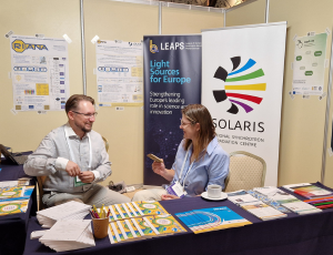
In the end of 2017 a photoemission electron microscope had been successfully installed on SOLARIS PEEM/XAS beamline. For the first time in Poland synchrotron radiation was used to image the sample surface with chemical and magnetic resolution.
Several samples were under investigation during extensive XAS measurements. The most spectacular one was the permaloy sample on silicon substrate with Co spacer and Al capping aluminium layer (1nm Al / 2nm NiFe / 20nm Co / Si). On the lithographically modified sample surface one could observe nano- and micro- structures with different magnetic domains.
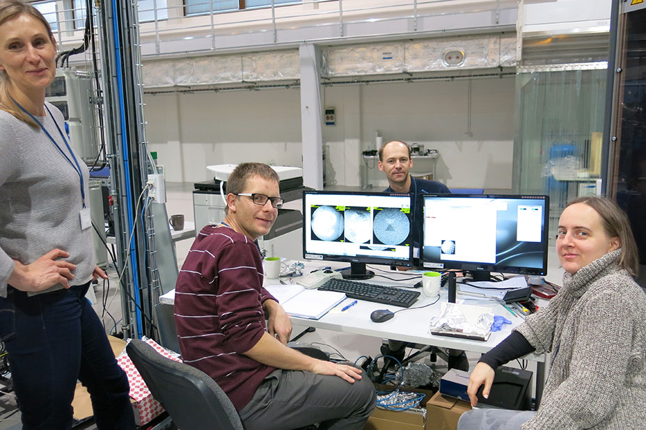
In the picture (from the left): Dorota Wilgocka-Ślęzak (IKiFP PAN), Michał Ślęzak (WFiIS AGH), Marcin Zając (NCPS SOLARIS), Joanna Stępień (ACMiN AGH).
 |
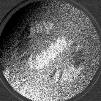 |
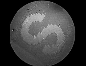
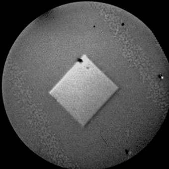
| S stands for SOLARIS. Above images of magnetic domains were obtained at the L3 Fe edge (706.8eV). On the left: XAS image acquired with linear polarization of synchrotron radiation. On the right: XMCD differential image of magnetic domains. The field of view is 75μm. | |
 |
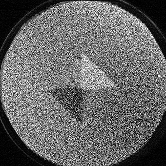 |
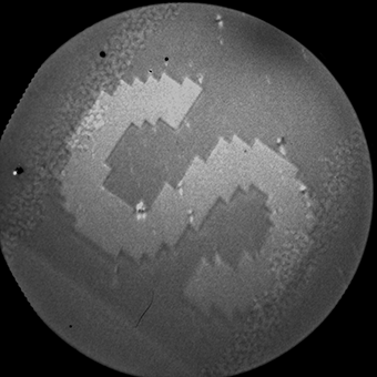
| Above images of magnetic domains were obtained at the L3 Fe edge (706.8eV). On the left: XAS image acquired with linear polarization of synchrotron radiation. On the right: XMCD differential image of the closed magnetic loop of Landau type domains. The field of view is 50 μm. | |
Modification date 09/04/2022
- Agnieszka Cudek
Recommended

Will artificial intelligence provide us with a cure for cancer or an elixir of youth? The iNEXT Conference.