
Scientific publications list
Scientists from the SOLARIS National Synchrotron Radiation Centre (Kraków, Poland), the University of Exeter (UK), Beckman Institute (the University of Illinois at Urbana-Champaign - USA), and the Institute of Nuclear Physics Polish Academy of Sciences (Kraków, Poland) performed research that will facilitate the rapid and automated diagnosis of esophageal cancer.
Dr. Tomasz Wróbel`s group focuses on cancer detection through a combination of Infrared Imaging (IR) and the use of Machine Learning (ML) algorithms. Thanks to this approach, it is possible to develop an effective model, which will allow histopathologists to confirm the diseased area automatically and in a much shorter time.
Infrared Imaging (IR), which will soon also be available on the newly built beamline at SOLARIS, has found widespread use in biomedical research over the last couple of decades and is currently being introduced into clinical diagnostics.
The wealth of biochemical information provided by this method is contained in spectra, describing unique characteristics of sample to absorb infrared light of different wavelengths - part of IR spectrum is often called a “molecular fingerprint”. It also allows us to identify hundreds of chemical compounds. Combining multidimensional data from IR Imaging with Machine Learning (ML) algorithms led to the development of a completely new field focused on cancer detection – explains Dr. Tomasz Wróbel.
Using samples coming from healthy and cancerous patients (originally diagnosed by a trained histopathologist), ML algorithm searches through spectra to find and extract features characteristic for a given tissue type and therefore, creates a model. This model is later applicable for identification of future and unknown samples.
Such full automation, which does not require staining of samples, is a great support for histopathologists and allows them to detect inflammation and pathology in the body faster and much more effectively.
To be suitable for clinical application, a potential method needs to fit into a specific timescale. In case of IR Imaging, this issue can be aided by Quantum Cascade Laser-based (QCL) infrared spectroscopic imaging that provides a more rapid method (minutes per patient) than conventional Fourier transform infrared imaging (FT-IR). However, apart from speed, there are plenty of other aspects, for example sample preparation, that needs examination and further optimization – adds Dr. Wróbel
This work considers a few factors, having crucial impact on the final work-flow of the proposed diagnostic approach. First of all, how effective will the model translation be to a faster modality of QCL microscopes. FT-IR measurements provide full spectral information, which is not always needed in full for model creation. On the other hand, QCL based Imaging allows focusing only on selected spectral regions. Defining a small subset of spectral frequencies, which is most important for tissue types discrimination, provides a basis to create reliable classification model, giving at the same time such needed time advantage. Results show that even after major reduction of spectral information, models achieve sensitivity of around 95%.
Apart from the mentioned project, Dr. Wróbel’s group is also working on a Full Pancreatic Cancer classification model including cancer types and staging differentiation, inflammations and surgical margin detection with FT-IR Imaging. Such studies are one of the possible avenues of research that soon will be available on a newly constructed SOLAIR beamline at National Synchrotron Radiation Center SOLARIS.
The research team included scientists: Danuta Liberda - Jagiellonian University, NSRC SOLARIS; Michael Hermes - University of Exeter; Paulina Kozioł - Institute of Nuclear Physics - Polish Academy of Sciences; Nick Stone - University of Exeter; Tomasz Wróbel (corresponding author) - Jagiellonian University, NSRC SOLARIS and Beckman Institute, University of Illinois at Urbana-Champaign.
The complete publication Translation of FT-IR imaging esophagus histopathological model to a fast QCL modality can be found in Journal of Biophotonics.
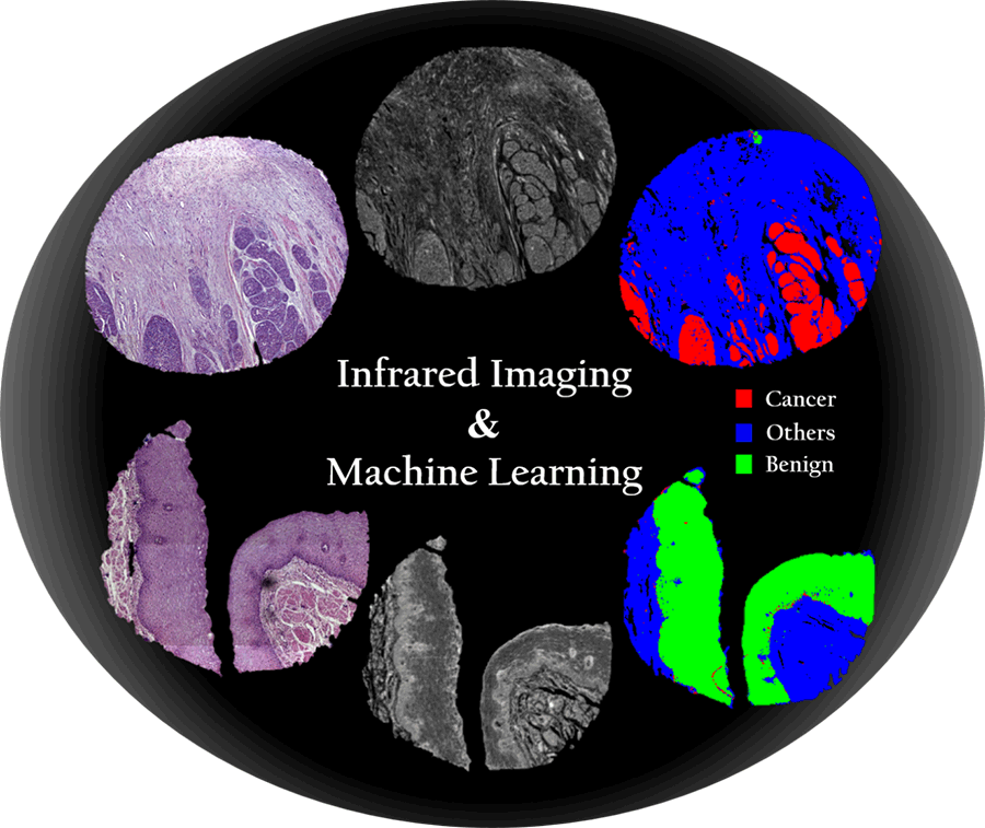
The above graphic shows two esophageal biopsies: the top of the graphic contains a biopsy taken from a patient suffering from esophageal cancer, the bottom of the graphic contains a biopsy taken from a healthy patient. In the left part of the graphic, microscopic images of the mentioned biopsies are visible after the H&E staining (Hematoxylin and Eosin) (in this image of the stained biopsy, the histopathologist visually assigns tissue types), in the middle of the graphic, biopsy images obtained using infrared imaging are visible, the right part of the graphic presents a histological picture of a biopsy obtained after assigning tissues and structures to three classes (cancer, other, benign) by Machine Learning (ML).
Red - cancer
Blue - other
Green – benign