
Scientific publications list

The prestigious journal ‘Analytical Chemistry’ (IF: 6.785) has recently published a paper written by the SOLARIS team and titled ”Macromolecular orientation in biological tissues using a four-polarization method in FT-IR imaging”.
Carried out in the SOLARIS Center in the framework of the Sonata grant funded by the National Science Center, the research project aims to develop a new infrared spectroscopy approach to analyzing biological samples. Its results give a detailed picture of the macromolecular organization e.g. of human tissue. The new four-polarization method can now be used, for the first time, to determine the collagen orientation in pancreatic tissue biopsies.
“The way that electromagnetic radiation is absorbed by the sample depends on its polarization, which is the basis of the new method. By carrying out measurements with four different linear polarizations and by employing a special mathematical model, we can unravel the macromolecular organization of the sample”, explains Paulina Kozioł, one of the authors of the paper. “This allows us to determine not only the direction of the main axis of collagen fibers, but also their level of organization”, she adds.
“FT-IR imaging combines the wealth of information on the biochemical composition of the sample with the parameters of macromolecular orientation associated with the use of polarized light. All this data will be used at the subsequent stages of the project to enhance cancer diagnostic models and to better understand the microenvironment of the tumor. What happens in the surrounding area is often of decisive importance for the results of chemotherapy”, adds Tomasz Wróbel, PhD.
This approach will be offered on the SOLAIR line, which is currently being built.
Authors: Paulina Kozioł (1,2); Danuta Liberda (1,2), Wojciech M. Kwiatek (2); Tomasz P. Wróbel (1,2)
1 – SOLARIS National Synchrotron Radiation Center, Jagiellonian University
2 – Institute of Nuclear Physics, Polish Academy of Sciences
Full text of the article is available here.
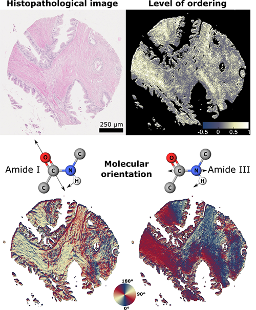
A biopsy specimen rich in collagen fibers, seen as pink, cylindrical areas in the histopathological image. The regions exhibit a high degree of organization and characteristic (nearly perpendicular) protein group (amide I and III) vibration directionality.