Scientific publications list
The FT-IR spectroscopic imaging in combination with machine learning algorithms has a great potential as a support for histopathologists in distinguishing types of tissues and cancer diagnosis. The introduction of the method to clinical practice is limited by the relatively high cost of sample substrates and the long preparation and measurement time. Depending on the selected measurement mode (radiation transmission or transflection), two types of sample carriers are particularly applicable.
In the case of transmission mode, the sample carriers are more expensive. However, the quality of the received signal is much better than in the case of the transflective mode. The sample preparation as well as the measurement times are the serious limitations because the samples, extracted from the patient, are placed in paraffin that is removed in a 24-hours deparaffinization process.
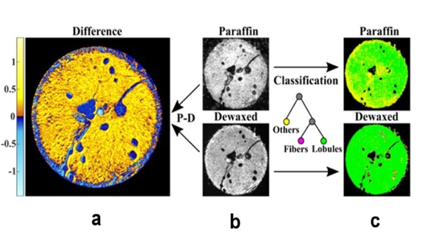
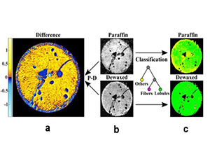
Taking into account the mentioned aspects, scientists from the SOLARIS National Synchrotron Radiation Center, the Institute of Nuclear Physics of the Polish Academy of Sciences and the AGH University of Science and Technology in the work "Influence of interference effects on the spectral quality and histological classification by FT-IR imaging in transflection geometry" analyzed the effects of signal distortions, obtained by FT-IR spectroscopy imaging (in transmission and transflection modes) of pancreatic tissues embedded in paraffin and after deparaffinization. The use of the machine learning algorithm allowed to evaluate the capability of this method in predicting the types of pancreatic tissues.
“The presented studies showed that the greatest signal distortion occurs in measurements in the transflection mode of paraffin-embedded pancreatic tissue samples, while the measurements of deparaffinized samples in the transmission mode showed the lowest signal distortion. Nevertheless, the accuracy of the classification was satisfactory in both measurement modes”, explains Danuta Liberda, one of the authors of the publication.
The results of the presented research performed within the PRELUDIUM grant, led by MSc Danuta Liberda carried out in our institution give hope for the possibility of implementing infrared imaging in clinical practice with the use of inexpensive substrates. This aspect will be further investigated using breast tissues within the grant mentioned above entitled “Improvement of histopathological classification based on chemical FT-IR imaging with data augmentation”.
Full version of the publication: here.