

Lista publikacji naukowych
Albicydyna jest niezwykle silnym antybiotykiem bakteriobójczym działającym na problematyczne bakterie Gram-ujemne, jednak jej mechanizm działania nie został poznany na poziomie molekularnym. Zespół współpracujących naukowców z Polski, Niemiec i Wielkiej Brytanii wykorzystał kriomikroskopię elektronową w SOLARIS do określenia struktury albicydyny związanej z kompleksem - gyrazą DNA z Escherichia coli wraz z fragmentem dwuniciowego DNA o długości 217 par zasad.
Toksyna peptydowa – albicydyna – jest produkowana przez roślinny bakteryjny patogen Xantomonas albilineans, który powoduje choroby liści u trzciny cukrowej. Cząsteczka ta jest wykorzystywana przez patogen do niszczenia chloroplastów roślin, umożliwiając jego rozprzestrzenianie się. Albicydyna hamuje replikację DNA w chloroplastach przez celowe hamowanie niezbędnej topoizomerazy DNA – gyrazy DNA, która jest również obecna we wszystkich bakteriach. Dzięki temu, albicydyna jest niezwykle silnym antybiotykiem bakteriobójczym działającym na problematyczne bakterie Gram-ujemne, jednak jej mechanizm działania nie został poznany na poziomie molekularnym. Zespół współpracujących naukowców z Polski, Niemiec i Wielkiej Brytanii wykorzystał kriomikroskopię elektronową w SOLARIS do określenia struktury albicydyny związanej z kompleksem - gyrazą DNA z Escherichia coli wraz z fragmentem dwuniciowego DNA o długości 217 par zasad.
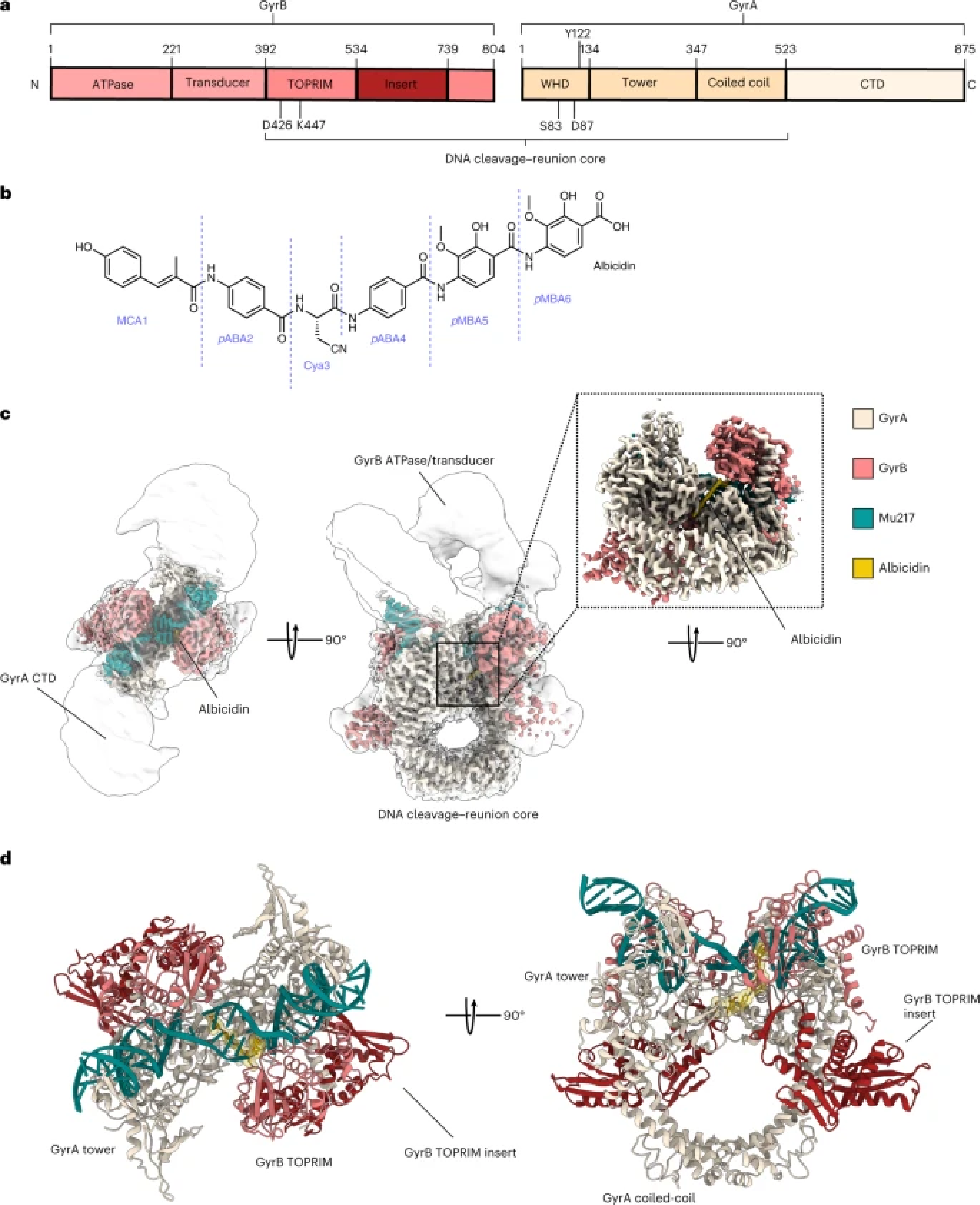
Rysunek 1. a, Scheme of GyrA and GyrB domains. The DNA cleavage–reunion complex is shown, along with the catalytic residue (Tyr122) and residues involved in quinolone resistance (GyrB Lys447 and Asp426 and GyrA Ser83 and Asp87). A consistent colour code is used throughout the manuscript: beige, GyrA; coral, GyrB. b, Chemical structure of albicidin. c, An overview of the Gyr–Mu217–albicidin cryo-EM map depicted as an overlay of two different contour level maps. Low-resolution contour (white) illustrates the position of GyrA CTDs and GyrB ATPase domains. High-resolution core part, including albicidin (in the zoomed-in image), is coloured according to the scheme in a: coral, GyrB; beige, GyrA; teal, DNA; yellow, albicidin. d, Cartoon representation of the overall model.
Odkrycie:
Struktura o wysokiej rozdzielczości (2.6 Å) kompleksu Gyr-Mu217-albicydyna ukazuje kompleks złożony z dwóch podjednostek GyrA i dwóch podjednostek GyrB, DNA o długości 217 par zasad owiniętym wokół gyrazy, które jest pocięte tworząc wiązanie kowalencyjne między DNA a katalityczną tyrozyną w białku. Albicydyna jest dobrze widoczna w otrzymanej gęstości, a jej pozycja wykorzystuje unikalny, podwójny sposób wiązania, gdzie N-końcowa część albicydyny interkaluje między pocięte DNA, a C-końcowa część klinuje się między dwiema podjednostkami GyrA blokując ich ruch i postęp cyklu katalitycznego enzymu. Dodatkowo, przeprowadzono badania stukturalno-aktywnościowe oraz określono struktury syntetycznej pochodnej albicydyny o wyższej aktywności, ulepszonej rozpuszczalności oraz aktywnej w badaniach na modelach zwierzęcych.
Otrzymane struktury pokazały, że N-końcowa część albicydyny może być podstawiona przez płaskie aromatyczne grupy ze znanym powinowactwem do DNA takie jak chinolina czy naftalen. Co interesujące, aktywność albicydyn nie była hamowana przez mutacje występujące w gyrazie DNA odpowiedzialne za oporność na fluorochinolony, dzięki czemu albicydyny tworzą nową grupę trucizn topoizomeraz i są atrakcyjnym potencjalnym lekiem.
Zrozumienie struktury wiązania pozwoli teraz na dalsze wykorzystanie nowej kieszeni wiążącej i wprowadzenie większej liczby modyfikacji albicydyny w celu poprawienia jej skuteczności i właściwości farmakologicznych.
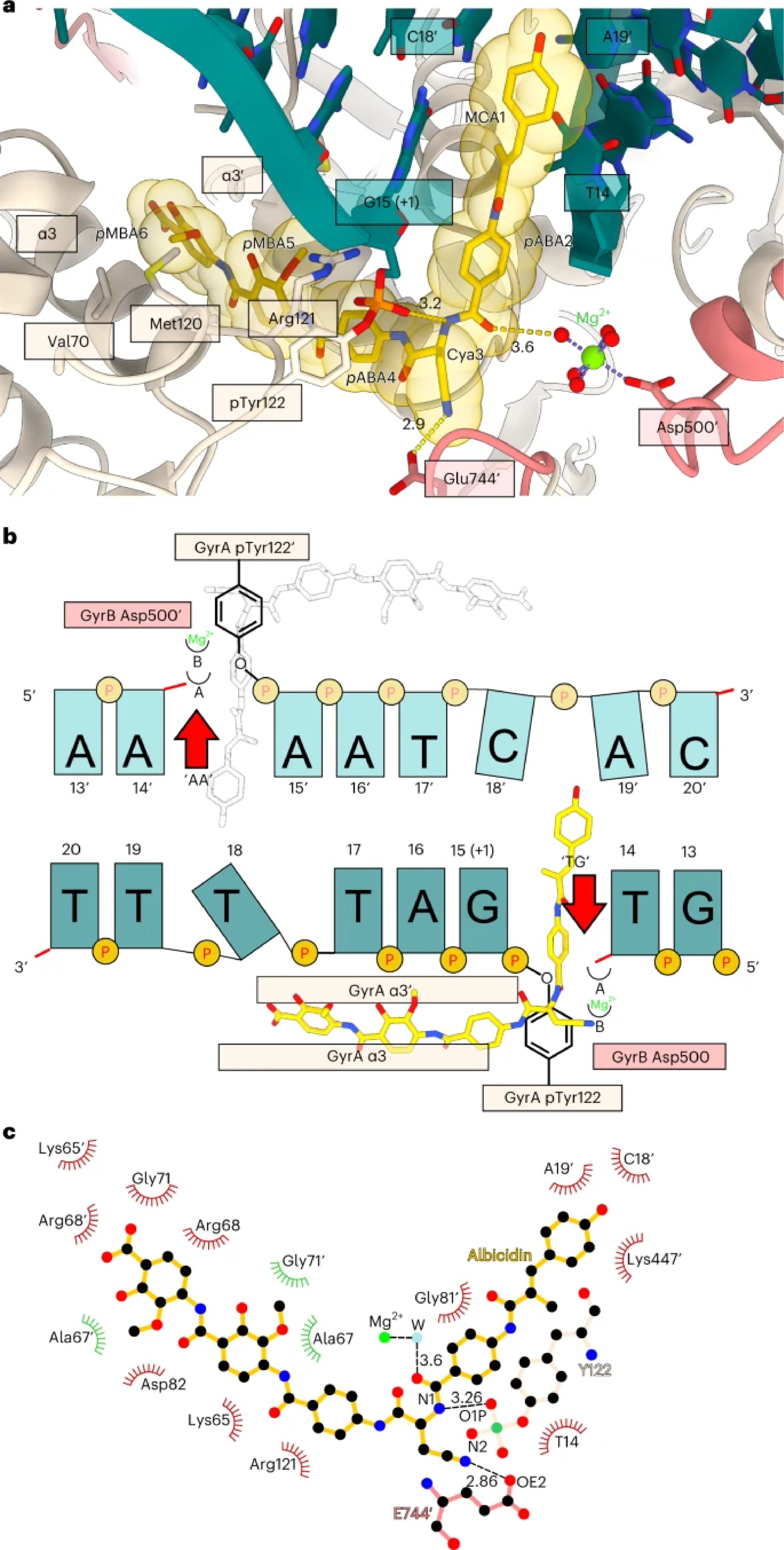
Rysunek 2. a, Enlarged view of the albicidin-binding site in the Gyr–Mu217–albicidin structure. Gyrase is represented as a cartoon, and albicidin as a stick representation. Van der Waals radii for albicidin atoms are shown as transparent yellow spheres. Two opposing GyrA helices α3 and α3′ at the dimer interface (DNA gate) form one part of the binding pocket, while DNA bases form another part. Distances (Å) between the modelled metal ion water shell and GyrB Glu744′ to Cya3 of albicidin are indicated. b, Schematic of albicidin binding in the context of the Mu cleavage site. Two potential binding pockets next to the scission sites ‘TG’ and ‘AA’ are labelled by the red arrows. Albicidin position is depicted by the sticks model (yellow), with the grey image indicating the potential alternative orientation not observed in the Gyr–Mu217–albicidin data. Two metal-binding sites, A and B, are indicated as half-circles. c, A LigPlot76 two-dimensional diagram of the albicidin-binding site. Hydrogen bonds and lengths (<4 Å) are indicated with dashed lines and the non-bonding and hydrophobic interactions (<4 Å) are labelled by the red and green spiked arcs, respectively. W, water coordinated to the metal (presumed Mg2+) ion.
Napisane przez: Ghilarov Dmitry
Link do publikacji: