
The main parameter analysed in X-ray absorption spectroscopy is the absorption coefficient μ of the material as a function of the incident X-ray energy E.
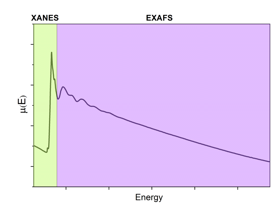
Figure 1. Dependence of the absorption coefficient from the photons energy (X-ray absorption spectrum μ(E)).
There are several modes of signal detection in case of determining μ(E).
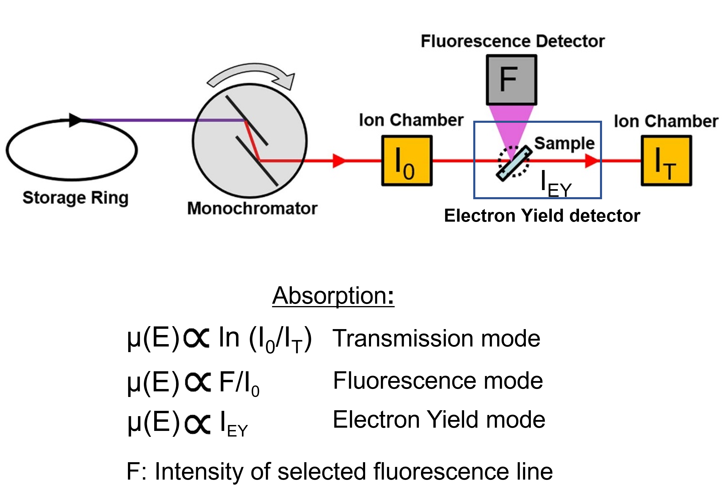
Figure 2. Measurment modes of XAS spectra.
Transmission mode XAS (Avaliable!)
In this mode the X-ray photons passing through the sample are directly detected. For each photon energy E, selected via Bragg axis rotation of the DCM, the absorption coefficient µ is calculated from the logarithmic ratio of the incoming intensity I0 and the intensity It transmitted through the sample:
µ~ln [ I0 (E) / It (E) ]
I0 and It are measured with ionization chambers, and a third ionization chamber is used to measure the intensity transmitted through a reference sample (e.g., metal foil for energy calibration of the DCM). The gases and the pressure in the ionization chambers are optimized for the energy range of interest.
The transmission mode is the simplest and the most accurate method for studying thin samples (with thickness roughly about 1–2.5 absorption lengths) with a high concentration of the absorber element (>3-5%). The thickness of the sample should be uniform on the scale of an absorption length. Also, the sample should be as uniform as possible over the width of the beam, without “pinholes”.
Fluorescence mode XAS (Avaliable!)
In this mode the intensity If of a selected X-ray emission line of the element of interest is measured by a fluorescence detector while scanning the incoming X-ray energy with the DCM. To minimize elastic scattering the detector is mounted perpendicularly to the incoming beam. Absorption spectra are then obtained by dividing If by the incoming intensity I0:
µ~ If (E) / I0 (E)
Since the concentration sensitivity in fluorescence mode is around 10−4% this measurement mode is chosen for diluted systems and biological samples, preferably with absorber concentrations below 3–5%.
Measurements in Total Electron Yield mode (Coming soon!) and Conversion Electron Yield mode.
Standard XAS measurements in transmission or fluorescence mode are bulk sensitive due to the high penetration depth of X-rays. Surface sensitivity can be achieved by detecting the electrons emitted from the surface of the sample or the electron current, i.e., Total Electron Yield (TEY). The probing depth can be changed and controlled by measuring Conversion Electron Yield (CEY), which is carried out in a gas environment, and the measured signal is due to electrons resulting from the ionization events initiated by high-energy electrons emitted from the sample.
Transmission mode XAS measurements require uniform and sufficiently thin samples to transmit X-rays, whereas for TEY there are no limitations with respect to thickness and uniformity. Additionally, in TEY mode the signal intensity is higher because all electrons emitted from the sample, including photoelectrons, Auger electrons and secondary electrons are collected. However, TEY detection mode is limited to the conductive samples. Surface sensitive measurements using XAS in TEY/CEY mode are very important in nanotechnology (multilayers, nanoparticles, nanowires, etc.) as well as for projects with industrial partners in the field of coating and adhesive technology.
Advanced measurements
Beamline staff is working on implementation of extensions for various in situ and operando experiments. In 2023 – 2024 binding of standard XAS measurements with complementary techniques (e.g., FTIR and Raman spectroscopy) are planned.
Please contact with beamline team in order to clarify the realization status of these upgrades.