The CIRI research line will ultimately be equipped with three end stations for imaging in micro- and nanometric spatial resolution. All end stations can use both a synchrotron source and a standard source (QCL or blackbody source). There is a possibility in the future to extend the line by a fourth end station.
The s-SNOM/AFM-IR microscope uses radiation in a wide spectral range. The atomic force microscope (AFM) is coupled to scanning near-field microscopy (sSNOM) and IR microscopy. The station is designed for microscopy with spatial resolution in the nanometric range.
Infrared spectroscopy exploits the fact that molecules absorb specific frequencies of light that are characteristic for their structure. The frequencies of the absorbed radiations match the frequencies of bond vibrations in the molecule. FT-IR microscopy uses this phenomenon to image the sample and obtain chemical information about it.
Depending on the detector used, it is possible to map the sample (collect successive single measurements) and image. The Bruker Hyperion 3000 microscope, which is the first end station of the CIRI line, has two detectors cooled with liquid nitrogen:
- MCT detector (mercury-cadmium-telluride detector): it is characterized by the highest sensitivity among the broadband mid-infrared detectors available on the market. MCT is a photon detector where electrons are excited directly by the absorbed radiation. MCT does not cover the full mid-infrared range because a minimum amount of energy is needed to excite the electron. In the case of our detector, the lower limit is approximately 800 cm-1. Mapping with the MCT detector is a point-by-point analysis of the sample. The study area during single measurements is limited by a mechanical aperture,
- FPA detector (Focal Plane Array): it is made of arrays of MCT detectors (64x64). Thanks to this type of construction, in a single measurement thousands of spectra are collected simultaneously, which makes the analysis much faster. The spatial resolution of the image is limited by the diffraction of infrared light to a few micrometers.
Experiments:
Depending on the type of sample their a few modes of measurement to choose from:
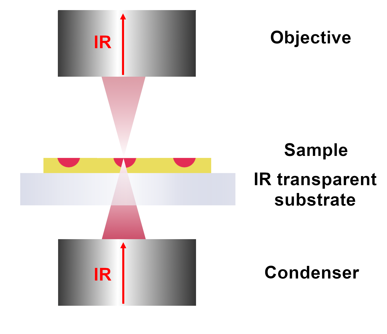
- Transmission mode: IR light comes out from the condenser, passes through the sample, where it is partially absorbed, and goes into the objective and then to the detector. The substrate on which the sample is deposited must be transparent to infrared light in the tested range. For the mid-infrared, the most common substrates are barium fluoride, germanium fluoride, or potassium bromide. The sample thickness should not exceed approximately 10 μm. The amount of absorbed radiation depends on the thickness of the sample, if it is too high, the signal obtained will be too weak for analysis.
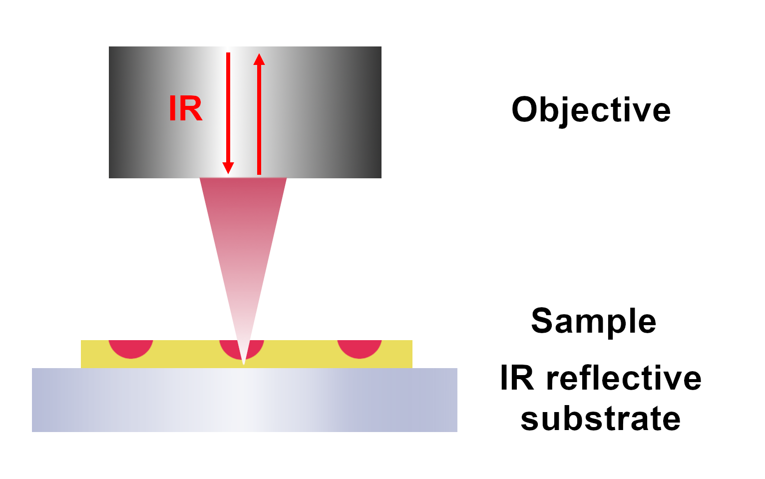
- Transflection mode: IR light comes out from the objective, passes through the sample, and is reflected on the substrate covered with a thin reflected, metallic layer. Next, IR light passes again through the sample and goes to the objective, and the detector. The transflection mode combines transmission and reflection techniques.
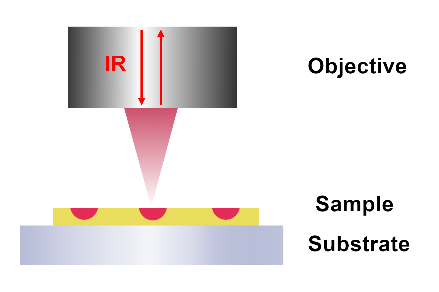
- Reflection mode: IR light comes out from the objective, is reflected on the sample surface, comes back to the objective, and goes to the detector. In the case of high roughness of the sample surface (> 1 μm), the signal quality is lower due to beam scattering on "unevenness".
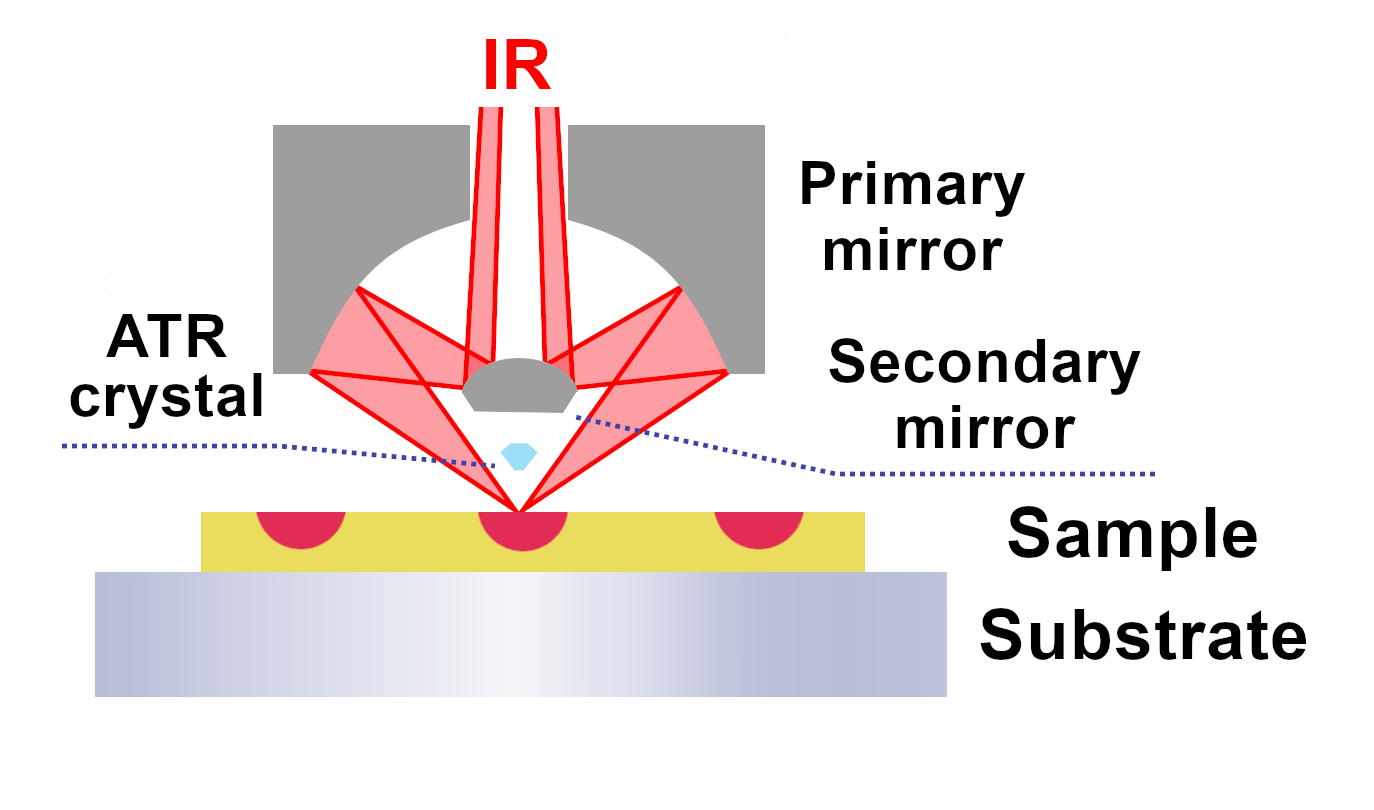
- ATR mode: exploits the attenuated total reflection effect. The measurement is performed after pressing a germanium crystal with a high refractive index to the sample surface. As a result of the interference of the incident wave and the reflected wave, a standing wave is formed, which propagates in the direction perpendicular to the boundary of the so-called evanescent wave. Since the penetration depth depends on the wavelength of the incident radiation, a correction to the intensity of the received signal is applied. The method allows you to record the spectrum with little preparation of samples.
The second end station of beamline CIRI is the Neapsec microscope. The sSNOM and AFM-IR measurements can be performed with nanometric spatial resolution below the diffraction limit of infrared light. The microscope construction is based on an atomic force microscope (AFM).
AFM-IR and sSNOM are complementary techniques. Depending on the characteristics of the sample and measurement needs, it is possible to select the appropriate technique or use both. The sSNOM scanning optical near-field microscopy enables imaging with spatial resolution at the nanoscale beyond the infrared diffraction limit. The detected signal is scattered light under the AFM probe. At the same time, information about the absorption of IR radiation, the optical properties, and the morphology of the sample is obtained. The nature of the signal depends on the optical properties of the sample and the material from which the AFM tip is made. Gold or silicon are used as reference, substrate materials. The sSNOM microscopy allows for obtaining a wide range of information about optical phenomena with a resolution of up to twenty nanometers. It allows for the exploration of the differences in optical properties of sample components on the nanoscale and allows for the study of light-matter interactions. The technique is dedicated to materials that strongly interact with light.
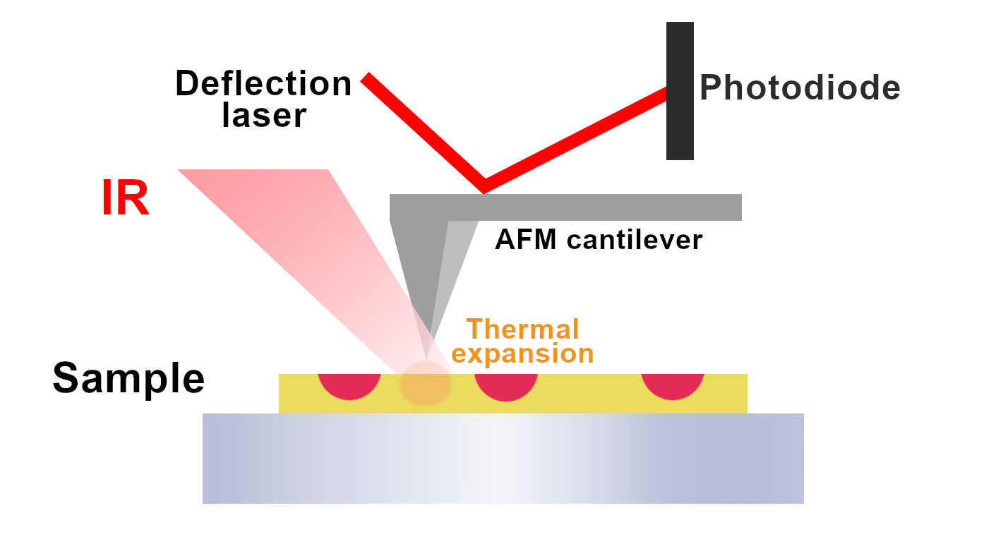
The AFM-IR technique is suggested when a full IR spectrum is desirable. The absorbed IR light is detected by the AFM tip which “reacts” to the thermal expansion of the sample. The thermal expansion effect of the material is mostly dependent on the sample's absorption coefficient for a given wavelength and is largely independent of the optical properties of the sample and probe material. The method is more dedicated to testing soft materials, characterized by a higher coefficient of thermal expansion.
The IR radiation source may be a synchrotron providing an ultra-wide range of wavelengths from near (NIR) to far (FIR) infrared or a laser source enabling narrowband imaging. The quantum cascade laser (QCL) allows for fast imaging at specific frequencies.
A summary of both techniques:
- sSNOM: the measured signal is a scattered IR light, which goes to the nitrogen-cooled MCT detector. The IR light is focused on the AFM tip and interacts with the sample surface. sSNOM measures the amplitude and phase of the scattered light in the near field. At the same time, information about the chemical and optical properties of the sample is obtained. The character of the signal depends on the wavelength, the material's absorption coefficient, and the refractive index. The metal AFM tip limits the area analyzed while amplifying the electric field at the same time. sSNOM provides high spatial resolution (nanometers) and surface sensitivity. Measurements are made on reflective substrates, usually, gold or silicon plated.

- AFM-IR: light absorption by the sample is measured using the phenomenon of photothermal expansion. The expansion of the sample under the AFM tip. Changes in the cantilever vibration frequency are measured by a laser. The technique allows for obtaining the full spectroscopic spectrum of the sample. It is often used in research on polymers, composites, and materials of biological origin - cells, tissues, and protein aggregates.
List of Neaspec AFM-IR technologies:
tapping AFM-IR+: tapping mode AFM-IR imaging & spectroscopy.
PTE: legacy contact mode PTE technique suitable for thick samples.
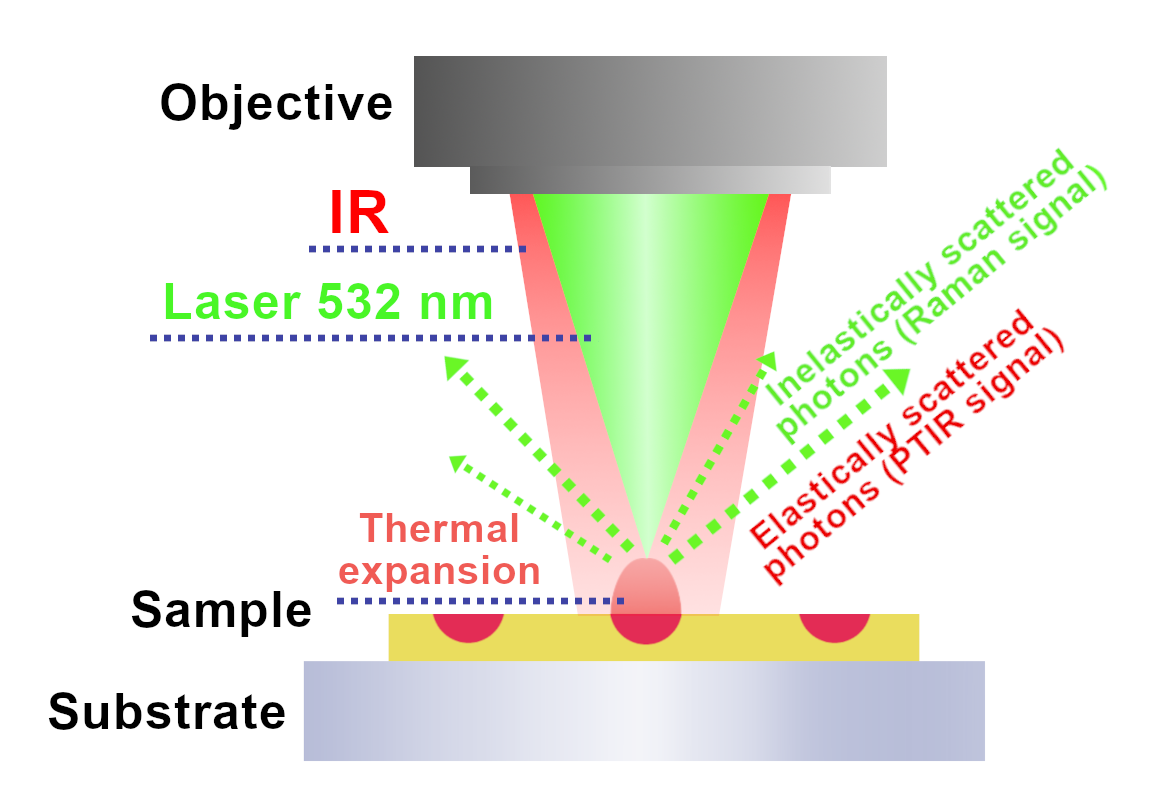
The O-PTIR microscope is the third end station of the CIRI beamline. This is the second technology developed in recent years to obtain infrared images with spatial resolution below the IR light diffraction limit. The microscope has two light sources - a laser with a wavelength in the visible range (532 nm) and an infrared radiation source - a pulsed, tunable QCL / OPO laser or a synchrotron source with a wide wavelength range.
The sample is simultaneously illuminated with green laser light and infrared light. If the frequency of the IR wave matches the frequency of the molecule’s vibrations, absorption occurs, and as a result, the photothermal expansion of the sample in the place of illumination. The expansion changes the intensity of the elastically scattered light (Rayleigh scattering) of the green laser. The obtained signal is used to generate the spectroscopic infrared spectrum. The photothermal response depends on many parameters related to the sample, including the thermal conductivity, the heat capacity and density of the absorbing molecules, as well as the power of the probe (visible laser) and the power of the infrared source. The spatial resolution is limited mainly by the wavelength of the laser acting as the probe (532 nm).
Besides the elastic scattering of photons used for IR detection, there is also inelastic scattering (Raman effect), i.e. the formation of photons with altered energy. As an effect, the Stokes and anti-Stokes lines appear in the spectra symmetrically on both sides of the Rayleigh line, with respectively reduced and increased frequencies. The O-PTIR method allows, thanks to the use of a green laser, to obtain both infrared and Raman signals at the same time.
Samples for O-PTIR measurements do not need any special preparation. The form of a thin film isn’t required. The method is non-destructive, without contact with the sample. However, it should be noted that the use of too high laser power may damage some types of samples (biological, polymers).