
The concept of the beamline DEMETER (Dual Microscopy and Electron Spectroscopy Beamline) assumes the coexistence of two measurement branches, in which the research end-stations is:
- the PEEM microscope (photoemission electron microscope),
- the STXM microscope (scanning transmission X-ray microscope).
The PEEM end-station consists of an Elmitec LEEM/PEEM III with a Kirkpatrick-Baez refocusing mirror pair focusing the beam down to 12 x 36 µm2 (footprint on the sample).
Photemission electron microscopy (PEEM) is a specialized technique for advanced studies of morphology, electronic and chemical properties, and the magnetic structure of surfaces, thin-films and interfaces with spatial resolution at the nanometer scale. PEEM microscope belong to the family of so-called cathode lens microscopy, where the sample is a part of the objective – cathode that emits electrons. In a standard PEEM setup a UV lamp acts as a photon source. However, Elliptically Polarized Undulator installed at the DEMETER beamline variable polarization soft x-rays are delivered to PEEM.
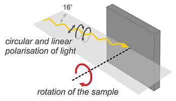
Photon are impinging the sample 16o with respect to the sample surface, what is schematically shown in Figure. The sample illuminated photon beam emits electrons which are further transmitted to the imaging chamber, creating the recorded image. The elastically scattered electrons together with others emitted from the sample are transferred to the imaging column through transfer lens. At the end of imaging column, the multichanelplate MCP detector is installed which amplify the numbers of electrons that hit the phosphorus screen. In order to create the microscopic image of the sample, the column is equipped with a system of electromagnetic lenses: a transfer lens that transfers electrons from the beam separator to the column and indirectly corrects the beam, an intermediate lens that selects the operating mode between real and diffractive imaging, and two projection lenses that magnify the image. Just behind the lenses, the multichanelplate MCP detector act as an image intensifier.
The final element of the column is the screen on which the resulting image is displayed. It is recorded through a CCD camera mounted outside the microscope. The entire system is additionally equipped with a hemispherical analyzer placed in the imaging column. Thus, polarized light sources in combination with the analyzer offer a wide range of other surface sensitive measurement techniques such as X-ray Absorption Spectroscopy, X-ray Photoelectron Spectroscopy, and Resolution Photoelectron Spectroscopy. angular ARPES (Angle Resolved Photoemission Spectroscopy) or X-ray Photoelectron Diffraction XPD.
In addition to chemical contrast, fully variable polarization enables magnetic imaging with X-ray magnetic circular and linear dichroism (XMCD and XMLD) of ferromagnetic and antiferromagnetic materials.

Rys. 1 (a) PEEM diagran, (b) Elmitec III PEEM in SOLARIS Centre.
The energy resolution of the microscope is defined by both the resolution of energy analyzer (below 0.15 eV) and resolution of the bemline (normally is negligible in comparison to resolution of energy analyzer). In the X-ray absorption mode, the energy resolution is determined only by the resolving power of the beam E / ΔE, which can be adjusted up to 8000 over the entire photon energy range.
In addition to x-ray absorption spectroscopy, in PEEM microscope we can perform microscopic X-ray photoemission spectroscopy (μ-XPS) in which a micrometric-sized area is selected. The dispersion plane of the analyzer is then projected onto the detector, whereby the photoemission spectrum from the selected area on the sample is recorded in one shot enabling real-time microspectroscopy. The spectral width is approximately 10 eV with a resolution less than 0.15 eV.
X-ray photoelectron diffraction (XPD) is another working mode that can be applied at PEEM microscope. It allows for imaging several Brillouin zones simultaneously for the selected kinetic energy of electrons. This method is especially useful for studying the dispersion of the valence band in micrometer size regions.
The standard operation conditions: the sample is at a high potential of 15 kV and 2 mm from the objective lens. Limited field mode (10 kV at the same working distance) is available for special samples and / or sample holders with an increased risk of discharge, such as insulating samples, surface electrodes, degassing surfaces, etc.
X-PEEM method and equipment
In a photoemission microscope with soft X-rays as an excitation source, XAS, XMCD and XPS spectroscopies are implemented with a lateral resolution of tens of nanometers. In this way, surface imaging with chemical, electronic and magnetic sensitivity is realized.
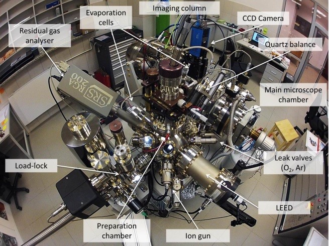
The station is equipped with an Elmitec photoemission microscope PEEM III with energy analyser. Samples are mounted on dedicated holders.
A preparation chamber, directly attached to the microscope, is as a standard equipped with:
- vacuum load-lock;
- LEED/AES Spectrometer;
- metal (Fe, Co, Au, Ni) and MgO vapour sources controlled with a quartz crystal balance;
- broad beam ion gun (Ar);
- O2 dosing;
- typical single crystalline substrates.
Sample requirements
- UHV compatible;
- flat;
- should not charge under X-ray illumination;
- diameter < 14 mm, height < 3 mm.
Imaging conditions
- probing depth of a few nanometers;
- image acquisition time from msec to several tens of minutes;
- field of view 5 μm - 150 μm;
- sample temperature 100 K - 1200 K (300 K - 2000 K in preparation chamber);
- maximum imaging pressure ~ 1*10-6 mbar.
Measurement geometry
The incidence angle of the synchrotron beam is fixed to the 16 degrees. The sample can be rotated over 360 degrees about the axis normal to the sample surface (azimuthal).
The second branch of the DEMETER beamline is ended with Scanning Transmission X-ray Microscope (STXM).
Scanning transmission X-ray microscopy (STXM) is a method to obtain a microscopic image of the raster-scanned sample by detecting the transmission intensity of the focused X-rays.
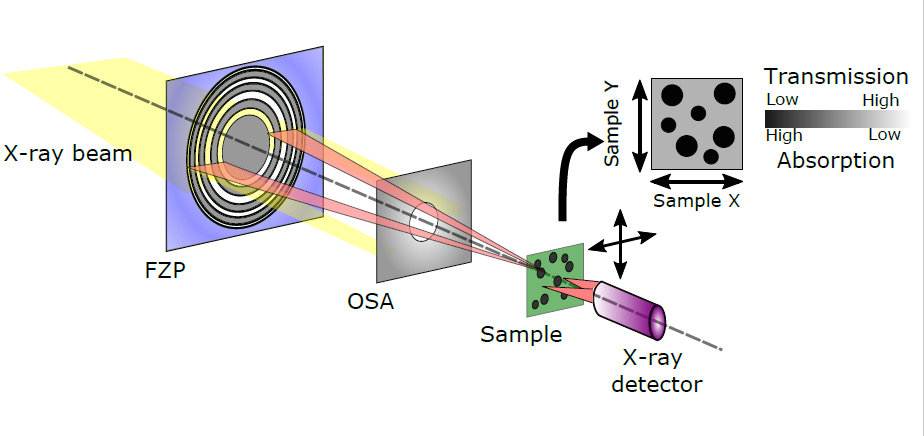
One of the main elements of the STXM microscope is the zone plate known as Fresnel zone plate (FZP). The FZP is focusing a monochromatic beam of photons delivered by the synchrotron. After passing through the Fresnel lens, the photon beam passes through the order-sorting aperture (OSA), used to select the diffraction fringes of the specific order. An unfocused zero-order beam is stopped by the OSA aperture and the central stop (CS) on the FZP lens. The sample is located at the focal point of the Fresnel lens. The focal distance from the FZP to the sample is typically several mms. The sample position is raster scanned with piezoelectric scanner, and simultaneously monitored with laser interferometric sensors.
Fast and low-noise avalanche photodiode (APD) is often used to detect the transmitted X-rays. In the case of weak signals, a photomultiplier tube (PMT), that can count single photons is better solution. PMT counts the photons created by excitation of a specific scintillator with X-rays transmitted through the sample. Advanced digital control electronics, with FPGA circuits, are used to realize a fast scan by selecting the dwell time at each pixel.
In principle, all the optics should be in vacuum, however, the ultra-high vacuum is not necessary. For samples that cannot be in vacuum, a pure helium (He) atmosphere can be provided in the microscope chamber. Other way is to sealed the sample between two membranes (made a so-called "sandwich"). The STXM chamber containing the elements from FZP to the detector is separated from the synchrotron beam by a thin Si3N4 silicon nitride membrane window. This allows a quick replacement of the sample and feasibility for in situ environments.
The samples measured by the STXM should not be too thick (this results in no transmitted signals) or too thin (no significant absorption). Ideally, the sample should have an optical density OD = ln (Io / I) ≈ 1 at the target photon energy range. For this purpose, the specimens for STXM are often prepared by dispersing the particles onto Si3N4 membranes or grid meshes with support films. Larger samples are usually sectioned by using microtomes or focused ion beam (FIB).
The lateral resolution, of the STXM microscope, is typically 20-100 nm and it is mainly determined by the diffraction limit of the lithographically produced Fresnel lenses (FZPs).
The most important measurement mode in STXM is the so-called "image stack" - a series of images are collected as a function of photon energy to obtain a dataset with space (XY) and energy (E) dimensions. A local absorption spectrum can be obtained from the arbitrary region of interest (ROI) at the image. This allows detailed analysis of the chemical composition of a measured sample.
Polarization-dependent experiments such as XMCD and LD are possible as with the bulk spectroscopy. In comparison with PEEM microscope were dichroic measurements are performed using bulk samples, in STXM only thin layers can be studied. Magnetic domains are obtained by calculating the asymmetry A = (IL - IR) / (IL + IR) from the images taken at two opposite circular polarizations (left and right).
Application of soft X-ray absorption on the nanoscale
The scanning transmission X-ray microscope (STXM) was designed and built at the Solaris synchrotron in close collaboration with PhD. Tolek Tyliszczak (Advanced Light Source, Berkeley, USA). The main mode of operation of the STXM microscope is X-ray absorption spectroscopy XAS, often referred to as NEXAFS (Near Edge X-Ray Absorption Fine Structure). This spectroscopy provides elemental and chemical specificity, as well as sensitivity to polarisation effects related to the magnetic and crystal structure of materials using XMCD and XMLD effects. Using a scanning X-ray microscope, chemical information can be obtained on a scale of tens of nanometres to several millimetres.
Measurement technique
The standard working mode at STXM microscope is transmission. However, but thick samples, or samples with small concentration of a given element, X-ray fluorescence yield (XRF) mode is available. Please note, that in fluorescence mode the resolution is lower than in transmission mode and the sample takes much longer to scan.Measurement geometry
Samples are scanned in a plane perpendicular to the beam in a focal point of a focused beam. In special cases, samples can be rotated with respect to the photon beam (max. +/- 45º).
Sample requirements
The sample should be sufficiently thin to allow for minimum 5% transmission for X-rays in the required energy range.
Samples can be placed on Si3N4 membranes or TEM grids. Recommended membrane thickness is from 50 nm to 200 nm, depending on the X-ray energy range.
The vapour pressure of the samples have to be smaller than the minimum measurements pressure at the STXM chamber
The standard frame size is 5.0x5.0mm (square) or Ø=5.0mm (circular). Minimum frame size is 2.5x2.5mm or Ø=3.0mm, respectively. Maximum sample size is limited up to 19x10mm (HxV). In this case, it is possible to scan only 8 fields with a diameter 2.5mm. Sample thickness: max. 1.0mm.
In special cases, it is possible to use a different sample holder (contact and agree with the microscope operator is necessary)
See: the standard Al plate for sample holders.
Experimental conditions and environment
- Available gases: He, Ar, O2, N2, CO2.
- Measurements pressure range: from 1e-7 mbar to 1100mbar (the standard operating conditions at STXM is He atmosphere with pressure from 10 to 1000 mbar).
- Temperature range: at this moment room temperature (RT) measurements are only available.
- Maximum field of view: +/-50µm2 (piezo stage), +/-1,25mm2 (linear stage).
- Possibility of mounting up to 8 samples
Equipment currently available
- Available detectors: photo multiplier tube (PMT), avalanche photodiode (APD).
- Solid drift detector (SDD) - Amptek X123 Fast SSD detector with sensitivity for photons with energy down to about 200 eV (Expert Commissioning access only).
- Electrochemicall Cell system for liquid and gases from Hummingbird Scientific (Expert Commissioning access only).
During implementation
- Measurements using a controlled external magnetic field.
- Ability to control the measurement geometry (angle of incidence).
- Electrochemical cell system for suspensions or solids.
- Environmental cell system with heating option (RT-475K).
- 4Mpx sCMOS detector.