
Cryo-EM core system technical highlights
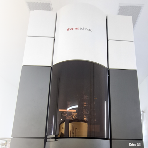
The state-of-the-art Krios G3i is equipped with a flexible three-condenser lens system and constant power lens optics, which in combination with a field emission gun (FEG) ensures very stable high-resolution measurements. The two direct electron detectors assure fluent workflow and high throughput of any single particle analysis (SPA) and cryo-electron tomography (cET) experiments. In its current configuration, the microscope captures up to 700 images per hour, making collection of obtaining a data set within a one-day measurement session possible. From SPA data, users routinely determine the spatial structures of macromolecules at resolutions in the range of 2-4 Å (current best 1.93 Å).
| Krios G3i Cryo-EM state-of-the-art Cryo-Electron Microscope |
|
|---|---|
|
 |
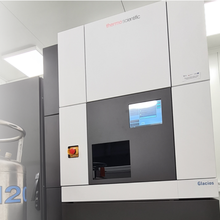
Glacios, the second microscope, is used for both data collection and sample screening. It is equipped with a direct electron detector dedicated to SPA and cET techniques, and a dedicated CMOS camera enabling microcrystal electron diffraction (MicroED) experiments. The microscope is capable of recording up to 400 images per hour, and the resulting density maps reach up to 3 Å for moderate-size particles (≥400 kDa) and 4-8 Å for small particles (<200 kDa).
| Glacios Cryo-EM high-end data collection & sample screening Cryo-Electron Microscope |
|
|---|---|
|
 |
Microscope Access
SOLARIS Cryo-EM Facility offers two pathways of access to cryo-microscopes:- The Krios G3i microscope is an integral part of SOLARIS DUO system, where one can submit a research application for a 2.5-days experimental shift. Applications can be submitted twice a year in the spring and autumn calls. All applications are assessed by the international scientific committee. The best applications are granted time on the Krios G3i microscope. For information on the feasibility of the experiment please contact cryo-EM specialists: dr Michał Rawski at michal.rawski@uj.edu.pl, dr eng. Paulina Indyka at paulina.indyka@uj.edu.pl, dr Marcin Jaciuk at marcin.jaciuk@uj.edu.pl.
- The second way of accessing the cryo-electron microscope at SOLARIS Cryo-EM Facility is paid access, allowing academic researchers to freeze and screen samples utilizing the Glacios microscope. The service not only offers the collection of preliminary data required for Krios G3i application, but also offers structure reconstruction packages for industrial and commercial customers (available techniques). For more information about the access and prices please contact dr Piotr Ciochoń, Industry Liaison Officer at industry.solaris@uj.edu.pl.